Study Notes
Corkin (1997)
- Level:
- AS, A-Level, IB
- Board:
- AQA, Edexcel, OCR, IB, Eduqas, WJEC
Last updated 22 Mar 2021
Effect of hippocampal damage on memory.
Background information: Corkin had known H.M. since 1962, during which time he had never recognised her from one visit to another. Corkin and her colleagues used MRI scanning in 1992 and 1993 (written up in their 1997 article) to determine if Scoville’s estimated lesioning of H.M.’s temporal medial lobe area had been as he stated (see the diagram under Scoville and Milner), and whether this could be sufficient to have resulted in the drastic memory loss suffered by H.M.
Aim: To investigate the extent of the hippocampal and medial temporal lobe damage to H.M.’s brain and to determine whether this could be sufficient to have resulted in the drastic memory loss suffered by H.M.
Method: One MRI scan was conducted on H.M. in 1992 and one in 1993. Before the 1992 scan, H.M. completed an IQ test and a memory test. The IQ test showed that he had normal intelligence, but the memory test showed his memory quotient (MQ) was 37 points lower than his IQ and showed he had severe amnesia.
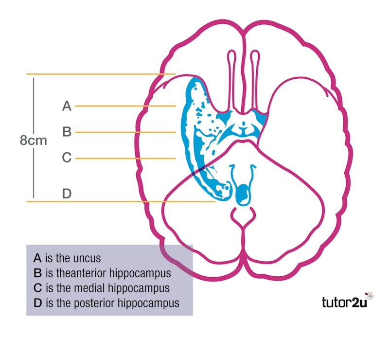
Results: Both scans showed that the lesioning (also called ablation or cutting) of H.M.’s brain was 3cm less than Scoville had estimated. It therefore did not extend as far into the posterior hippocampal region as he thought, although there was surrounding damage, as stated, to the uncus and the amygdala. Approximately 50% of the posterior hippocampus on each side remained, but this had shrunk considerably on the right side. Corkin et al. believe this could be due to both the removal of the rest of the hippocampus, and also to the drugs and continuing (though much reduced) epileptic seizures.
Conclusions: The small amount of normal hippocampus remaining in the left temporal lobe was not sufficient to support normal memory. Therefore, this study demonstrates the importance of the hippocampus and the temporal medial lobe area for memory.Evaluation:
There is not much to criticise with this study. Corkin had interviewed H.M. extensively over the years, and took care to ensure that the MRI caused no trouble to H.M., who had three non-magnetic clips inserted in his brain by Scoville in 1953, and which had they been magnetic would have meant an MRI was not advised. However, ethically there are some questions. It was Brenda Milner, the psychologist associated long-term with H.M. who gave the permission for Corkin to scan H.M.’s brain. It is not clear if she was the appointed responsible adult legally able to do this. H.M., even if he gave permission himself, would not have remembered it, so there are issues with informed consent and right to withdraw, although anonymity was maintained until after his death.
For More Study Notes…
To keep up-to-date with the tutor2u Psychology team, follow us on Twitter @tutor2uPsych, Facebook AQA / OCR / Edexcel / Student or subscribe to the Psychology Daily Digest and get new content delivered to your inbox!
You might also like
Scoville and Milner (1957)
Study Notes
Fink et al. (1996)
Study Notes
Maguire et al. (2000)
Study Notes